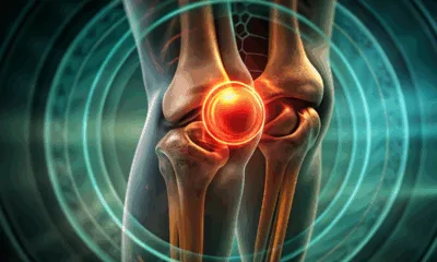
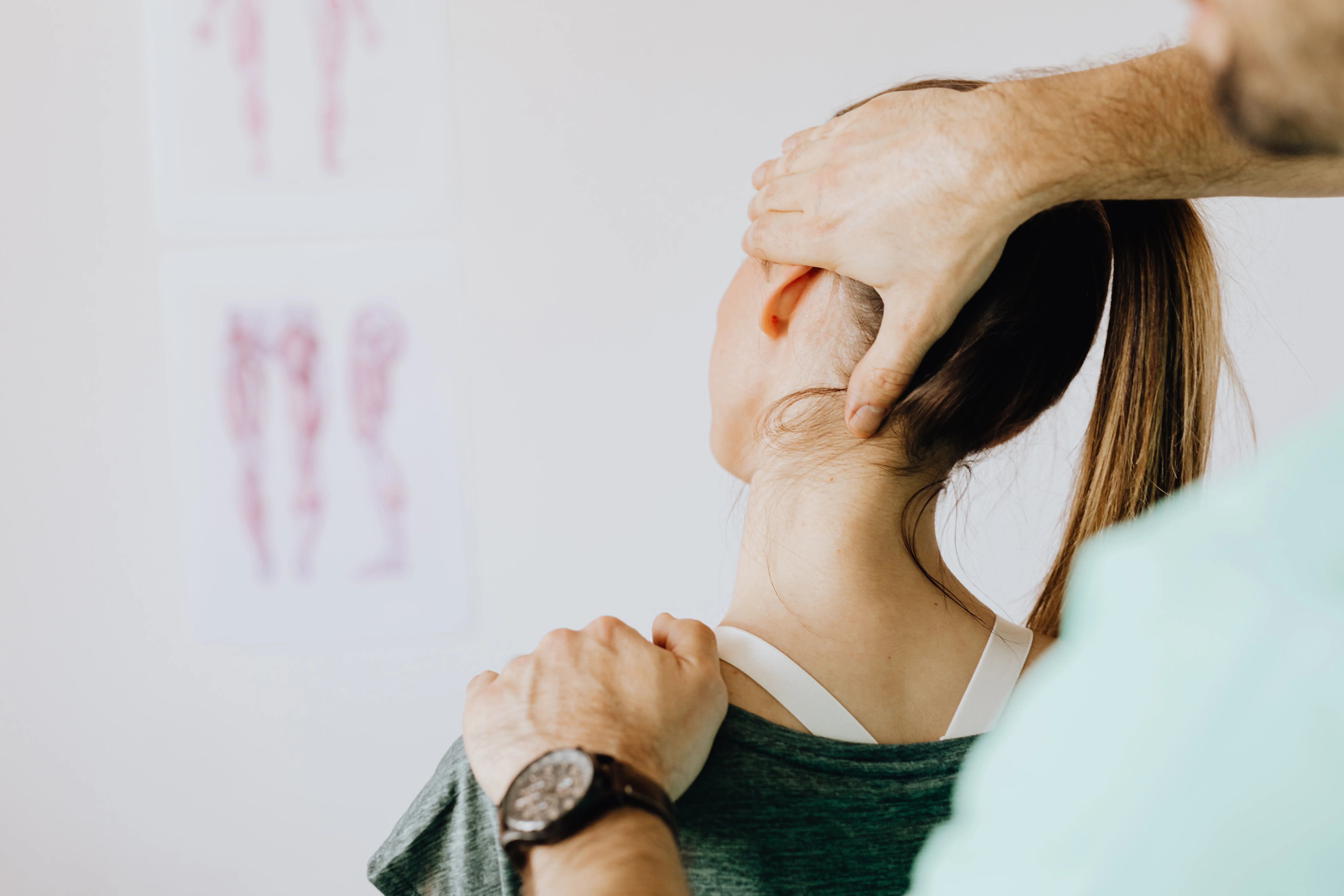
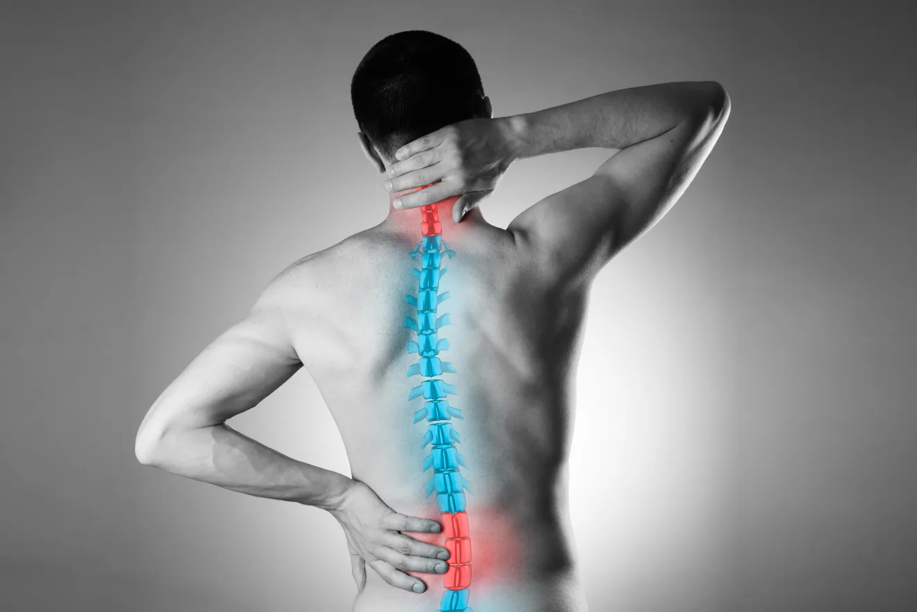
Osteopenia, or low bone mineral density, increases fracture risk. It is a precursor of osteoporosis – a more severe form of bone mineral loss that makes the bones weak, thin, and highly prone to fractures. Both are diagnosed with a bone density scan.
Osteopenia, or low bone mineral density, increases fracture risk. It is a precursor of osteoporosis – a more severe form of bone mineral loss that makes the bones weak, thin, and highly prone to fractures. Both are diagnosed with a bone density scan.
Bone density scan
Bone density scan, or dual-energy X-ray absorptiometry (DEXA/DXA) scan, is a quick, non-invasive x-ray imaging that measures bone strength.
Two different energy X-ray beams are passed through bones. By measuring how much is absorbed by the bone and soft tissue, the test calculates the bone mineral density (BMD).
Recommendations for bone density tests
Currently, there is no clear consensus on the optimal timing for bone density tests. In some Asian countries, it is recommended mainly based on WHO guidelines, for:
Females aged 65 years or older
Postmenopausal women younger than 65 years with risk of fracture
Perimenopausal women with clinical risk factors for osteoporosis
The 2025 Asia-Pacific Consensus for Management of Osteoporosis in Men recommends screening of all men aged 50 and above.
Younger people with an increased risk of bone loss may need the test too. Ask your doctor when you need this test and how often. Health conditions and medications that can reduce the BMD include:
Some autoimmune diseases
Use of tobacco
Drinking >3 servings of alcohol each day
Vitamin D deficiency
Use of corticosteroids
Some types of:
Cancer medicines
Immunosuppressants
Seizure medications
Medicines that treat acid refluxes
Use of heparin, a blood thinner
Getting your bone density tested
You may not need to change your usual routine before a bone density scan. Eat and drink before the test. Avoid taking calcium supplements at least 24 h before the test. Tell your doctor which vitamins and supplements you take. They will let you know which ones you can or cannot take before the scan.
The test usually lasts 30 minutes. It may be slightly shorter or longer depending on how many bones need scanning.
You will lie down on a special X-ray table.
A technician will position your body.
There will be no pain as no injection is needed.
The technician will pass a scanning arm over your body and take pictures of your bones.
In the image, the bones will appear white while fat, muscles and other soft tissue will look like dark shadows in the background.
How the results are interpreted
The test generates a number that is compared against one of two groups, depending on the patient. For postmenopausal women and men ≥50 years, the T-score is used. It compares them against an average, healthy 30-year-old adult. For premenopausal women and men <50 years, the Z-score is used. It compares them to people of their own age and gender.
Compared to the reference group, a standard deviation of -1 or more is considered normal, of -1 to -2.5 is a sign of osteopenia, and of -2.5 or less indicates osteoporosis.
It is possible to have different bone density scores in different bones depending on how metabolically active a bone is. However, the diagnosis is based on the lowest score in the body. Hence, if you are diagnosed with osteoporosis in one area, you have it everywhere.
Conclusion
A bone density scan is a safe, quick and painless imaging procedure that helps early detection of bone loss. Its objective measurements of BMD helps clinicians assess fracture risk, monitor disease progression, and guide timely interventions.